
Worakrit Saiyasombat,[a] Wachiraporn Kesorn,[b] Sirimongkhon Thukhammi,[b] Phiphob Naweephattana,[c] Bongkot Ouengwanarat,[d] Thanyada Rungrotmongkol,[c],[e] Anyanee Kamkaew,[d] Chutima Kuhakarn,[f] Onnicha Khaikate[b],*
[a] Institute of Nutrition, Mahidol University, Phutthamonthon, Nakhon Pathom 73170, Thailand
[b] Department of Industrial Chemistry, Faculty of Applied Science, King Mongkut’s University of Technology North Bangkok, Bangkok 10800, Thailand
[c] Center of Excellence in Biocatalyst and Sustainable Biotechnology, Department of Biochemistry, Faculty of Science, Chulalongkorn University, Bangkok 10330, Thailand
[d] School of Chemistry, Institute of Science, Suranaree University of Technology, Nakhon Ratchasima 30000, Thailand
[e] Program in Bioinformatics and Computational Biology, Graduate School, Chulalongkorn University, Bangkok 10330, Thailand
[f] Department of Chemistry and Center of Excellence for Innovation in Chemistry (PERCH-CIC), Faculty of Science, Mahidol University, Rama 6 Road, Bangkok 10400, Thailand
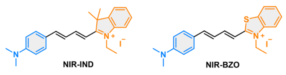
Intracellular viscosity is an important physicochemical parameter associated with cellular functions and disease progression. In the present work, two D–π–A hemicyanine-based near-infrared (NIR) fluorescent probes (NIR-IND and NIR-BZO) for sensitive detection of intracellular viscosity variations were developed. The probes feature a donor–π–acceptor framework that enables viscosity-responsive fluorescence through restricted intramolecular rotation. The 4-(dimethylamino)phenyl group serves as the electron donor, while two different electron-accepting groups (IND and BZO) are incorporated. Furthermore, two π-bonds were introduced to enhance fluorescence efficiency within the far-red to near-infrared (NIR) region (λ = 650–900 nm), enabling deeper tissue penetration. Both probes are readily synthesized and exhibit absorption and emission in far-red to NIR region. They exhibited moderate polarity sensitivity, pronounced viscosity-responsive NIR fluorescence properties, large Stokes shift (up to 162 nm), a broad pH stability range (3–13), high specificity, and strong photostability. They exhibit weak emission in low-viscosity environments and strong fluorescence enhancement under high-viscosity conditions. The probes are well suited for biological imaging. Confocal imaging revealed that NIR-IND and NIR-BZO were mainly localized in the mitochondria. Live-cell experiments demonstrate good cell permeability, low cytotoxicity, and effective visualization of intracellular viscosity changes, highlighting their potential for studying viscosity-related biological processes.
Saiyasombat, W.; Kesorn, W.; Thukhammi, S.; Naweephattana, P.; Ouengwanarat, B.; Rungrotmongkol, T.; Kamkaew, A.; Kuhakarn, C.; Khaikate, O. “Development of D–π–A hemicyanine based NIR fluorescent probes for sensitive detection of intracellular viscosity variations” J. Mol Struct. 2026, 1355, 145027. DOI: 10.1016/j.molstruc.2025.145027